


Before the ultrasound, before the calcium scan, before the lab draw, physicians made their living from inspection. Long before I could order a coronary CT, the cardiologists who trained me were reading the patient at the door: the earlobe, the eyelid, the ring around the iris, the color of the skin at the nape of the neck. Half of the finding was already in the room by the time the stethoscope came out.
Most of those clues live above the collarbone, which means your bathroom mirror sees them before I do. What follows are five things I look at on a patient’s face, and, roughly in the order you would notice them washing up in the morning, five things you can look at on yours.
None of the signs below is proof of heart disease. None of them is enough, by itself, to change your care. They are flags.
Think of the real drivers of cardiovascular risk as ingredients in a recipe. The familiar ones (high blood pressure, high cholesterol, smoking, diabetes, insulin resistance, a sedentary lifestyle, a poor diet, a strong family history of early heart disease, and chronic low-grade inflammation, which we sometimes measure with a high-sensitivity CRP test) are the ingredients that, when they collect in the same bowl, eventually bake the cake we are trying to avoid: a heart attack or a stroke. No single ingredient bakes the cake. The more of them accumulate in one person, the closer that person gets.
The five findings below are not the ingredients. They are visible clues on the outside of the bowl that an ingredient may already be inside. Early corneal arcus points at a lipid disorder. Acanthosis nigricans points at insulin resistance. Xanthelasma may point at how the body handles cholesterol at the tissue level. Persistently bleeding gums point at a chronic inflammatory state. A diagonal earlobe crease points at something we are still trying to understand.
None is a diagnosis. None is a reason to go request a specific test. Each is, at most, a reason to book a check-up and let your primary care physician decide what, if anything, is worth looking into.
Patient stories in this piece are composites; identifying details have been changed. The accompanying images were generated with AI software to illustrate composite clinical findings rather than photograph any specific patient.
1. A diagonal crease across your earlobe
The first time I noticed Frank’s sign on my own was in a 62-year-old accountant who had come in to the clinic for a routine heart checkup. No chest pain, walked three miles a day, took no medications. His earlobes carried a shallow, symmetric diagonal wrinkle I recognized only because a senior colleague had drawn it on a napkin for me years earlier during my training. The calcium score I later ordered from his age, family history, and lipids, not from the earlobe, came back elevated.
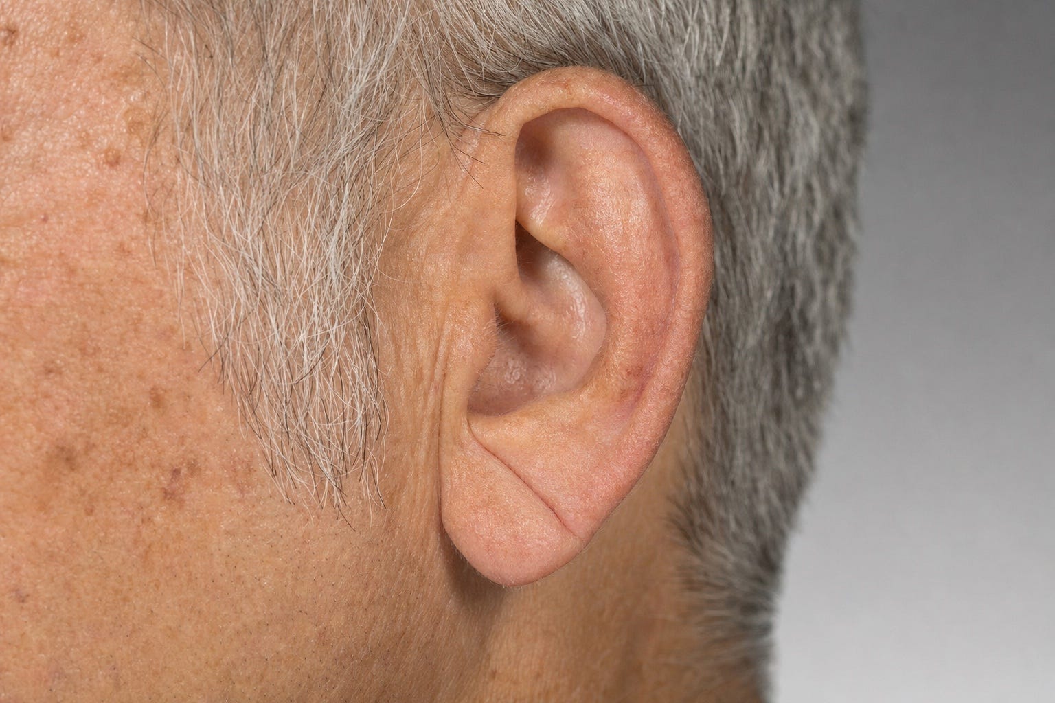
A diagonal crease running from the opening of the ear canal down and back toward the outer edge of the earlobe.
Turn your head slightly and look at your earlobe. A diagonal wrinkle that runs from the opening of the ear canal at about a 45-degree angle down and back toward the outer edge of the lobe is called Frank’s sign, after Sanders T. Frank, the American physician who first described it in 1973.
When you pool 12 studies and 5,000 people, people with the crease had roughly four-fold higher odds of coronary artery disease than people without it. That number is loud. It comes largely from patients who were already inside a cardiology setting (hospitals, catheterization labs, chest-pain clinics), where the prior probability of disease is high before the earlobe is even looked at.
Ask the same question in the general population and the answer shrinks. The Copenhagen City Heart Study followed nearly 11,000 adults for an average of 23 years. After adjusting for age, sex, cholesterol, blood pressure, diabetes, smoking, and family history, the crease was associated with about a 9 percent increase in ischemic-heart-disease risk. A separate diagnostic-accuracy review reached the same practical conclusion: the crease shifts the probability only slightly and should not, by itself, change what tests a person gets.
Bilateral creases, one on each ear, appear to carry a stronger association than a one-sided crease, at least in patients referred for coronary angiography.
Why would a wrinkle on the ear correlate with heart disease at all? No one is entirely sure. The leading hypothesis is that the earlobe sits at the far end of the ear’s blood supply, a small fatty flap with comparatively sparse vasculature and physiologically lower oxygen levels (autopsy series). That could make it prone to age-related, low-grade injury of the same type that is quietly damaging small vessels elsewhere in the body. Small biopsy studies have reported fragmentation of elastic fibers and thickening of small-vessel walls under creased earlobe skin, and one autopsy series found microscopic fibrosis and nerve changes at the base of the crease (Elliott et al.; autopsy series). These are real observations. They do not prove that ear-vessel injury and coronary injury are the same process, and part of what looks like small-vessel change on histology may simply be ordinary sun-related skin aging. Shared effects of aging, cardiometabolic risk factors, and connective-tissue biology are all still on the table.
The crease is not a diagnostic test. Plenty of people with it have clean coronaries, and plenty of people without it still develop heart disease. It is a flag on the door, not a note from the doctor. If you see one, especially on both ears, book the check-up you have been postponing.
2. Yellow plaques on your eyelids
A patient of mine once found her own xanthelasma in the visor mirror of her car, on the drive home from a lipid panel her physician had called normal. She sent me a photograph the next morning. Her LDL was in range; her HDL was not. The plaque did not send her to imaging by itself. It joined her family history, her HDL, and her age in a conversation that eventually did.
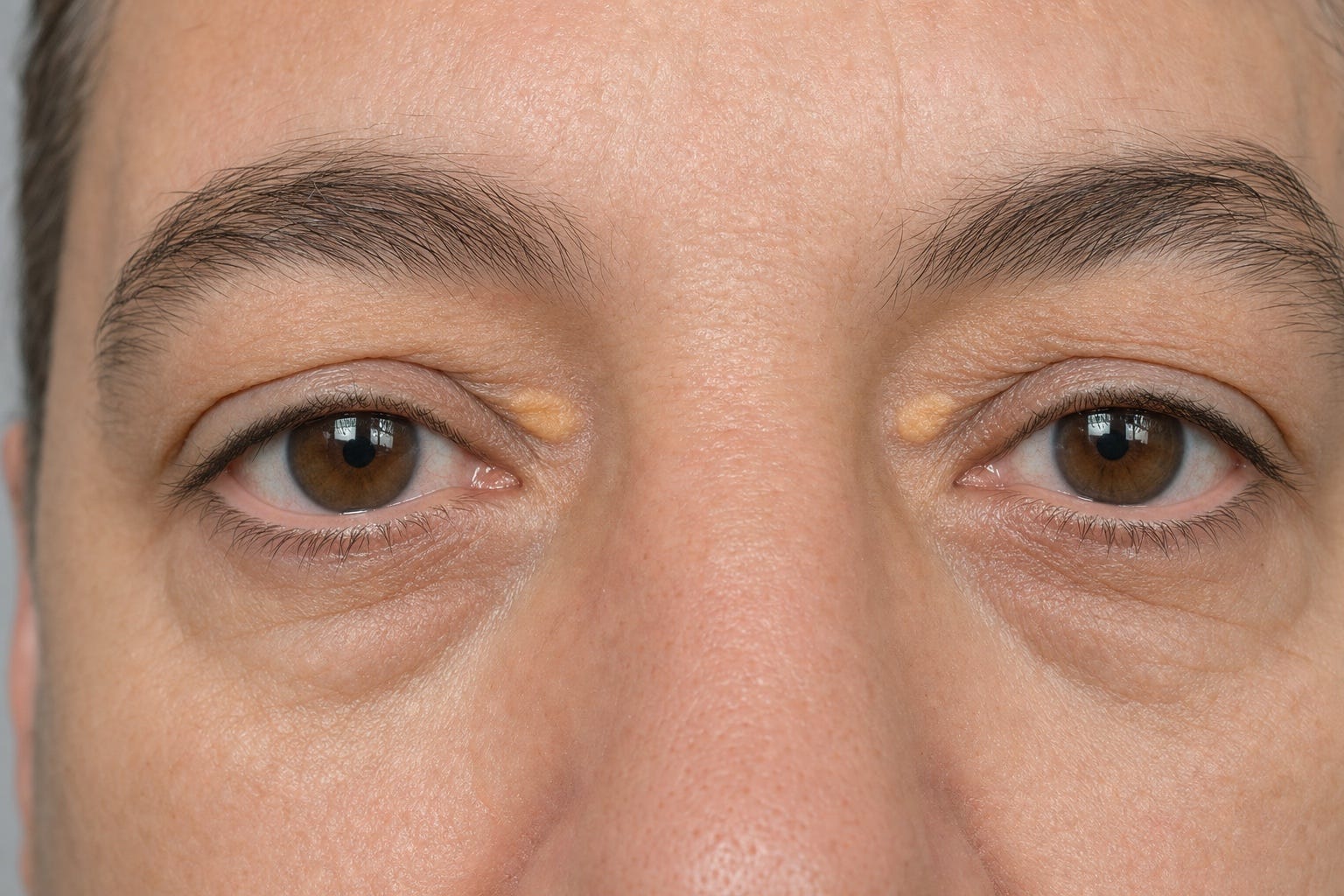
Xanthelasma: soft, yellowish plaques on the eyelids, most often near the inner corner by the bridge of the nose.
Xanthelasma are soft, yellowish, slightly raised patches on the upper or lower eyelids, most often near the inner corner by the bridge of the nose. Up close they look almost like a smear of yellow wax under the skin, which is close to what they are: macrophages that have swallowed cholesterol and settled where the skin is thinnest.
Their significance is easy to underestimate, because about half of people with xanthelasma do not have obviously high cholesterol on a routine lipid panel, though many still have subtler abnormalities, most often a low HDL. And yet the Copenhagen City Heart Study, nearly 13,000 people followed for up to 33 years, found that xanthelasma independently predicted heart attack, ischemic heart disease, and death, even after adjusting for cholesterol and triglycerides. The same study found no independent association with ischemic stroke.
My working interpretation is that xanthelasma may reflect something about how a particular person handles cholesterol at the tissue level that a standard lipid panel does not capture. Residual confounding and other pathways cannot be excluded, and this is still an area of active work.
One practical note. Cosmetic removal clears the plaque. It should not be assumed to change cardiovascular risk. The dermatologist takes the wax off the eyelid. Your primary care physician deals with what put it there.
If you see one, bring it up at your next visit, not your next cosmetic consult.
3. A pale ring around the colored part of your eye
A 38-year-old marathon runner sat across from me last winter and asked why his eyes looked older than his face. He had a soft grayish ring at the outer edge of each iris. His father had died of a heart attack at 47. His LDL, when we checked it, was 244. On the criteria we use for familial hypercholesterolemia, he scored as definite. Which meant the more important conversation was not really about him. It was about his brother, his sister, and his two children, who all needed a lipid panel of their own.
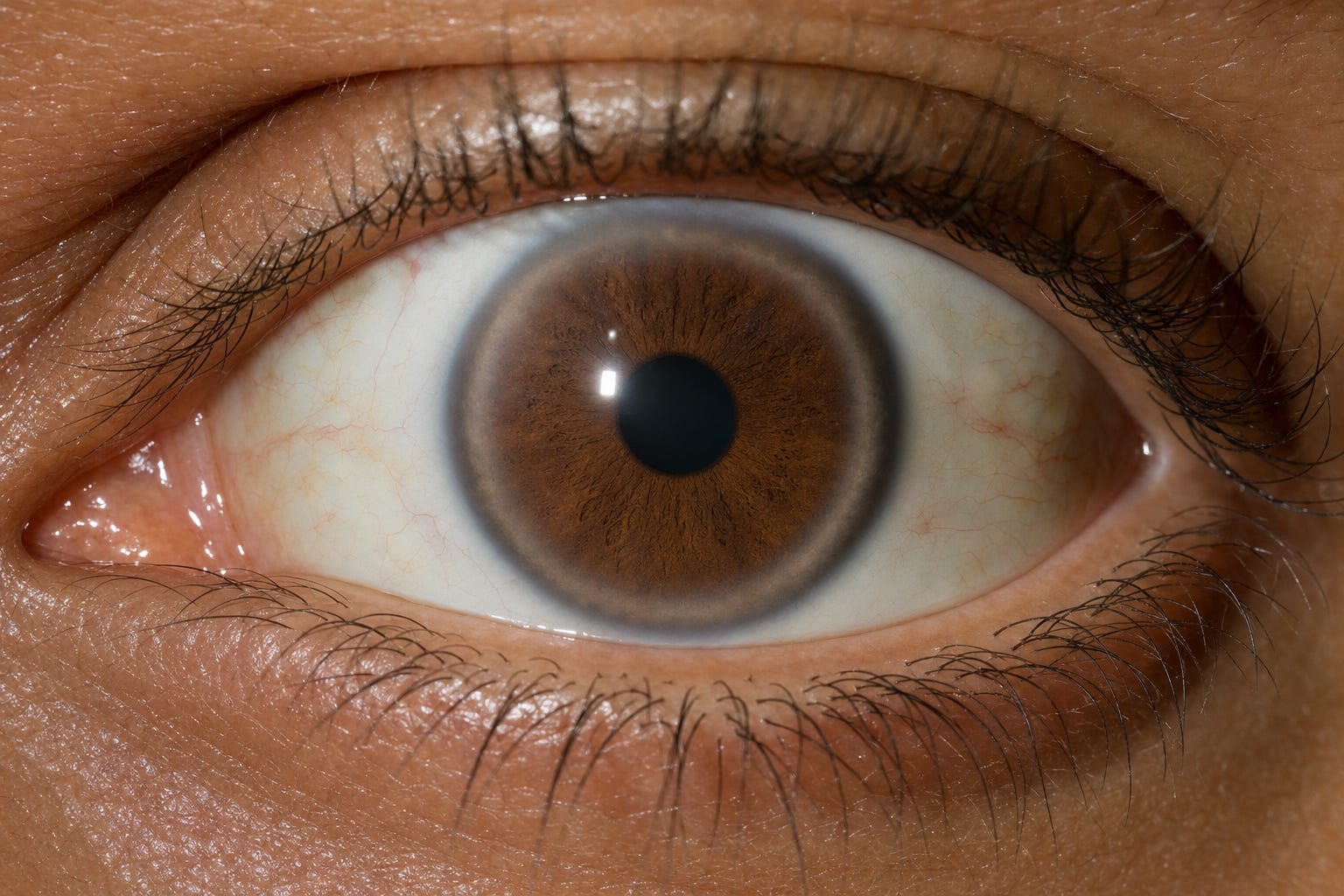
Corneal arcus: a grayish ring at the outer edge of the iris, formed by lipid depositing in the peripheral cornea.
Lean in close and look at the outer edge of the iris where it meets the white of the eye. A grayish-white, blue-white, or yellowish ring at that border is called corneal arcus. It is lipid, depositing in the peripheral cornea.
The interpretation depends almost entirely on age. In older adults, corneal arcus is largely an age-related change, and once you account for age the Framingham cohort found that arcus, by itself, no longer predicts cardiovascular disease.
Corneal arcus before age 45 is a different matter. It is a recognized criterion in the Dutch Lipid Clinic Network scoring system for familial hypercholesterolemia, an inherited condition where lifetime cholesterol exposure runs high from a young age and where treating early meaningfully changes outcomes. If a first-degree relative has been diagnosed, cascade screening (a lipid panel on parents, siblings, and children) is the highest-yield thing that finding can do. Arcus in anyone under 50 is still worth mentioning to your doctor as a possible clue to a lipid disorder, even outside the formal FH criteria.
One more thing to know. If the ring shows up on only one eye, that asymmetry has been associated with narrowing of the carotid artery on the opposite side. The mechanism is counterintuitive: the narrowed artery sits on the side of the eye without the ring, because reduced flow on that side keeps the lipid from depositing there. It is rare. It is worth mentioning specifically.
Under 50 and see either pattern? Treat it the way you would treat a xanthelasma. Book the visit.
4. Dark, velvety skin at the nape of your neck
A teenage patient once told me his mother kept scrubbing the back of his neck because she thought he was not showering. He was. The skin at the nape was velvety, brownish, and would not come off. His labs showed the picture of insulin resistance, and his BMI was in a range no one had thought to worry about. He was the exception, and that is part of why I remember him: most of the acanthosis I see is in people carrying extra weight. His story is the reminder that the skin finding can arrive first.
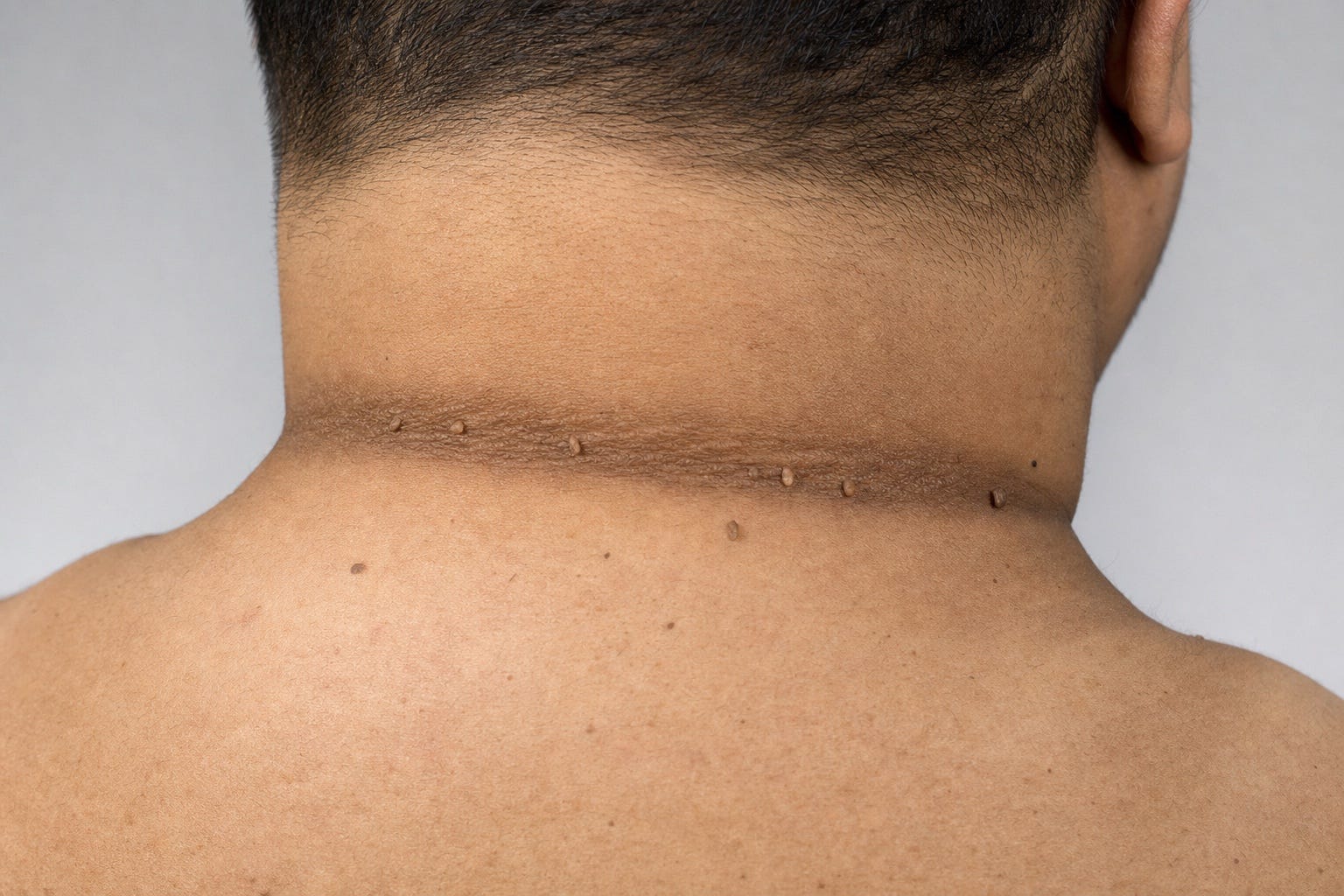
Acanthosis nigricans at the nape of the neck, with several small skin tags along the same crease.
Turn a bit and look at the back of your neck, at the folds under your jaw, and in your armpits. Brownish-to-black, thickened, velvety patches, the kind of finish that can make skin look dirty even after you have scrubbed it, are acanthosis nigricans. Small, soft, stalk-like skin tags (acrochordons) often keep them company along the same folds. On darker skin the color contrast is subtler, so look for the change in texture rather than the change in shade.
Both are visible clues to insulin resistance, the state in which the body has to make more and more insulin to keep blood sugar in range. Acanthosis nigricans is closely tied to hyperinsulinemia, and dermatologists have described it as a clinical stand-in for laboratory-measured insulin resistance, though the same review is careful to note that acanthosis is neither necessary nor sufficient for the diagnosis and that obesity itself is a stronger determinant. Skin tags are also nonspecific and occur commonly in people without any metabolic issue at all.
Insulin resistance is one of the central pathways linking abdominal weight gain to high blood pressure, fatty liver, unfavorable cholesterol patterns, and type 2 diabetes, most of which are relevant to the heart. In women, acanthosis nigricans can also be part of the picture of polycystic ovary syndrome.
One different scenario, worth naming plainly. Acanthosis that appears rapidly, spreads extensively, or is itchy in an adult can, rarely, be a paraneoplastic sign of an internal cancer, most commonly of the stomach or another abdominal organ. That pattern needs a same-week appointment, not a routine one.
In every version of this story, the destination is the same doctor. Only the timeline changes.
5. Your gums when you brush
The last thing most of us do at the mirror is brush our teeth, and the sink gives an honest verdict. Occasional bleeding after a change in floss technique is common and reversible. Persistent bleeding on brushing, gums that have receded up the tooth, and teeth that feel loose are the visible face of periodontal disease.
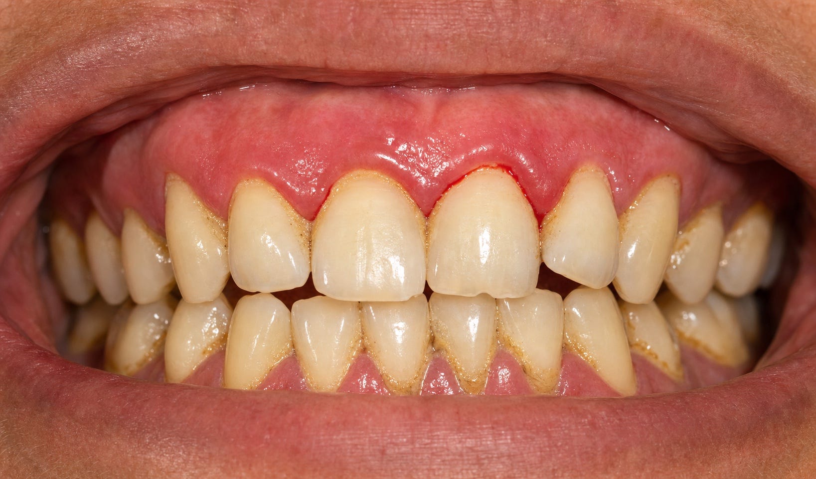
Inflamed, bleeding gum tissue at the margin between gum and tooth: the visible face of periodontal disease.
Of all five signs in this article, this is the one where it is easiest to overstate the case. Let me walk up the ladder honestly and stop where the evidence stops.
What is reasonably well established is that people with periodontitis have, on average, modestly higher levels of inflammatory markers like C-reactive protein and interleukin-6 than people with healthy gums. Treating gum disease is associated with a short-term reduction in CRP that is variable across trials, appears to fade by a year, and has not been shown in patients who already have cardiovascular disease. In the Tonetti randomized trial, intensive periodontal treatment improved endothelial function (how well small blood vessels relax and open) for several months.
What is not established is the final rung: that treating gums prevents heart attacks and strokes. No randomized trial has yet shown a reduction in hard cardiovascular events after periodontal treatment. Mendelian randomization studies, a genetic technique that can strengthen causal inference, have on the whole not supported a causal relationship between periodontitis and coronary disease or stroke. The American Heart Association’s 2025 update is explicit: gum disease is an associated risk marker for heart attack, stroke, atrial fibrillation, and heart failure, but a direct cause-and-effect relationship has not been proven, and shared drivers (smoking, diabetes, chronic inflammation, socioeconomic circumstances) may explain part of the overlap.
So where does that leave the mirror? Persistently bleeding gums are a visible clue that a chronic, low-grade inflammatory state may be present, and that some of the same drivers that damage gum tissue may also be acting on artery walls elsewhere. They are not proof of heart disease. Treating them is not a proven way to prevent it. But they are not “normal” either, and they belong on the list of reasons to see a dentist promptly and to mention the finding at your next primary care visit.
One safety note. Bleeding gums that are new, heavy, or accompanied by easy bruising, nosebleeds, or bleeding from other sites, especially in a patient on warfarin, a direct oral anticoagulant, or a strong antiplatelet regimen after a stent, need a same-week appointment rather than a routine one.
How to use these five
None of these signs, by itself, causes a heart attack. None of them is a causal risk factor, meaning something you could change and thereby change your risk. Each is a flag: a visible clue that a real risk factor may already be in the bowl.
A diagonal earlobe crease. The accumulated wear-and-tear of ordinary cardiovascular risk factors may already be at work. The underlying mechanism is still debated.
A xanthelasma. The body may be handling cholesterol in a way a routine lipid panel does not fully capture.
Corneal arcus before 45. A formal criterion for familial hypercholesterolemia. Arcus in anyone under 50 is a broader clue worth mentioning.
Acanthosis nigricans at the nape. A well-recognized clue to insulin resistance, with the caveats above.
Persistently bleeding gums. A clue to chronic low-grade inflammation, one of the modifiable contributors to cardiovascular risk over time, though treating the gums has not been shown to lower cardiac risk.
The real ingredients (high blood pressure, an unfavorable cholesterol pattern, smoking, diabetes, insulin resistance, a sedentary lifestyle, a poor diet, a strong family history of early heart disease, and chronic low-grade inflammation) are what actually bake the cake when enough of them collect in the same bowl. The five findings in this article are not enough, on their own, to bake anything.
They are the flags you can see for free every morning without a blood draw or a scan. Each one points to an ingredient a check-up can look for.
If you notice one, especially corneal arcus before 50, a xanthelasma at any age, new acanthosis nigricans, or long-standing bleeding gums, that is a legitimate reason to book a visit with your primary care physician, who can weigh it against the rest of your history and decide what, if anything, is worth testing.
The mirror does not diagnose anything. What it can do is show you a flag, and give you the chance to have that conversation a year earlier than you otherwise would.
In prevention, a year is often the whole margin.
If cardiovascular risk for women in and around menopause is what you came here for, that conversation lives at our sister publication, The Menopause Heart.
The Integrative Cardiologist is a free patient-education publication and is not a substitute for individualized medical advice. Nothing here constitutes a diagnosis or a recommendation to start, stop, or change any medication or treatment. If you notice a new or worsening physical finding, please speak with your own physician.